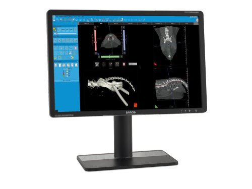
NNT: CUSTOMISABLE AND INTUITIVE SOFTWARE WITH DEDICATED VET INTERFACE
Doctors can access specific protocols and views according to body areas and diagnostic requirements; they can also program personal settings for re-use at a later time.
The advanced functions of the NNT software allow several medical specialties to be covered, and the special reconstruction windows respond to the different needs of each sector. All examinations are fully compatible in the DICOM format: they can be shared via NNT Viewer or printed in 1:1 scale.
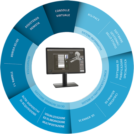
Complete connectivity
Excellent connectivity and integration with the modern systems adopted by NewTom. Workflow and clinical and diagnostic activities become much easier and highly performing.
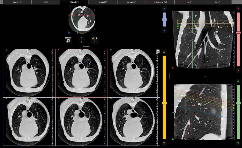
MULTIPLANAR REPORTING
The SLICE window creates datasets on the axial, sagittal or coronal planes, and allows the orientation and size of the volume to be changed on each of its axes, as well as to set the thickness of individual cross-sections. The advanced NNT functions facilitate reporting, with specific processing and sharing options for different medical specialties. Multiplanar analysis with personalised orientation allows body areas to be assessed from different angles.
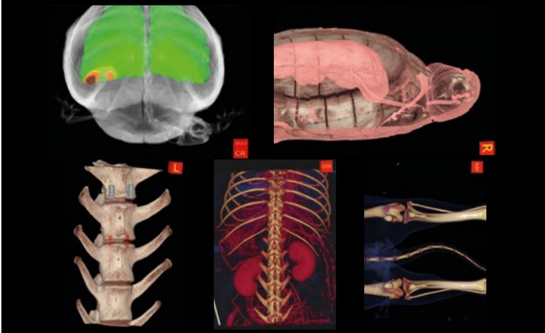
3D ANALYSIS
The simple 3D display interface makes communication with pet owners much easier, allowing the patient's condition to be made clear even to viewers who are not familiar with imaging reading. Separate or superimposed imaging of soft tissues and bone tissues can be chosen. 3D measurement, airway simulation and slicing tools are also available to obtain cross-sections of the volume of interest.
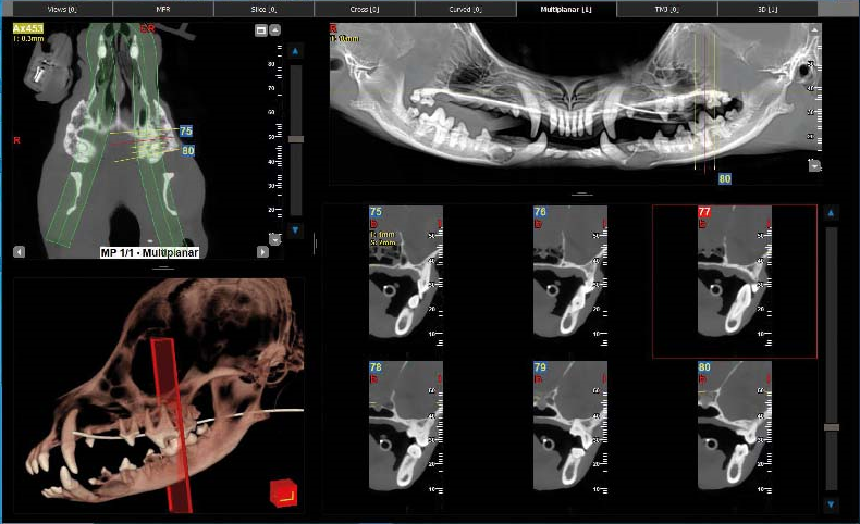
DENTAL PANORAMIC IMAGING
The dedicated interface for the study of dental arches generates cross sections and axial reconstructions, and produces images comparable to dental panoramic views with multiplanar reconstructions. It can also generate specific reconstructions for the coronal and sagittal planes. For all these images, thickness, brightness and contrast can be managed independently.
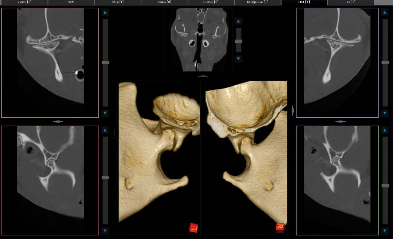
BILATERAL VIEWING
The NNT software has a dedicated window for bilateral imaging of bone structures, such as the temporomandibular joints and smaller joints. The viewing window shows the axial image in the middle and dedicated reconstructions for the left and right sides; in the bottom central area, the 3D renderings are shown.
