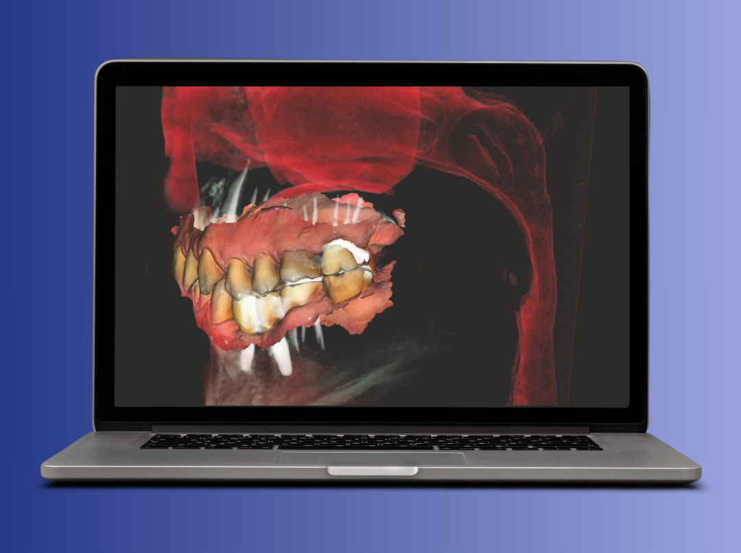
Diagnostic imaging is at the heart of modern veterinary radiology. Therefore, the integration of Cone Beam Computed Tomography (CBCT) technology in veterinary clinics and hospitals represents a true revolution. NEWTOM, a global pioneer in CBCT technology, brings its years of experience to the service of animal health with solutions that ensure ultra-high definition imaging and a patient-centered approach.
Diagnostic advantages of CBCT over traditional techniques
What exactly is CBCT technology and how does it work? Unlike traditional 2D radiography that provides a flat view of structures, CBCT uses a cone-shaped X-ray beam to capture a full volume of the patient in a single rotation. The result is a wealth of three-dimensional (3D) data that provides the veterinarian with an unprecedented diagnostic capability.
What are the diagnostic advantages of CBCT over traditional techniques?
- Superior anatomical detail
3D visualization, with resolutions up to 90 µm, as in the case of the NEWTOM 7G, is essential for diagnosing complex pathologies such as microfractures, joint lesions, periodontal disease, or middle/ inner ear pathologies that are often not visible with standard radiography. - Reduction of overlap artifacts
Complex structures do not overlap, improving image clarity, especially in areas such as the skull and spine. - Application versatility
With devices such as NEWTOM 7G, diagnoses can be performed on all body areas, including the chest and abdomen, for patients of different sizes (up to 215 kg). - Patient safety
The proprietary SafeBeam™ system automatically adapts X-ray emission to the animal's tonnage, while the use of pulsed emission and variable collimation limits irradiation to only the area of clinical interest. In addition, reduced scan times minimize the duration of sedation required.
Advanced clinical applications for hospitals
Adoption of CBCT elevates diagnostic capabilities to specialized levels, enabling the most complex cases to be addressed:
- Tissue characterization
The Dual Energy system uses acquisition at two distinct radiant energies to obtain information on the chemical composition of tissues. This significantly improves the distinction between cortical bone, trabecular bone, and soft tissue, and is crucial for reducing metal artifacts. - Dynamic analysis
The CineX feature uses serial radiography to create video sequences that allow dynamic analysis of moving structures, such as joints. This is essential for the study of orthopedic and neurological problems, providing a unique functional view. - Complex diagnostics
Advanced protocols support investigations ranging from myelography to angiography, making CBCT an indispensable tool for comprehensive and reliable diagnoses.
To discover the full range of our CBCT devices and to access our veterinary radiology tutorials, visit https://www.newtom.it/en/veterinary-radiological-devices/ and https://www.newtom.it/en/radiological-technology/
.png)
.png)

.png)


