3 configurations to meet every clinical and diagnostic need
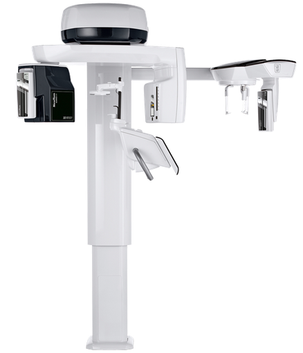
Unparalleled imaging
Amazing 3D FOV examinations and 2D protocols with the highest resolution images available on the market.
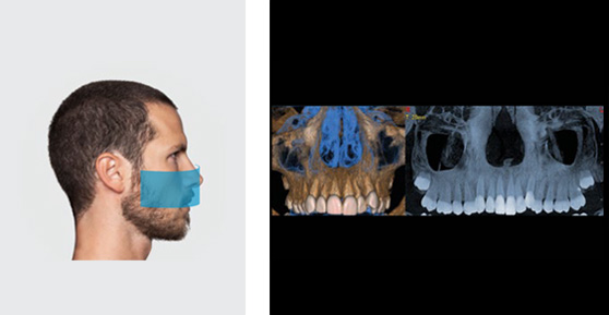
- 3D PRIME - 10 X 8
Highly suited to all dental diagnosis and implant-related needs. - 3D ADVANCED - 13 X 16
Broader vision: from maximum endodontic resolution to complete otorhinolaryngology examinations. - 3D PROFESSIONAL - 16 X 18
Full Head&Neck diagnostic performance to investigate the entire dental-maxillofacial district and cervical spine.
Essentially perfect
- General dentistry
- Implantology
- Endodontics
- Gnathology
- General orthodontics
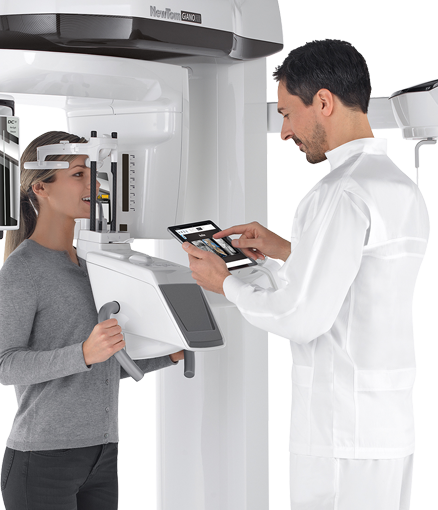
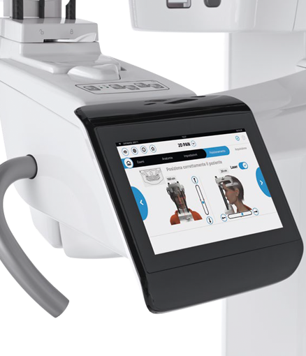
GiANO HR allows the user to always select the programme best suited to the clinical application with a few guided steps. Images of the complete dental arches can be obtained with a single scan or via ultra-high definition sectional acquisition in ECO-Dose mode (ultra-fast scanning).
Highly accurate, finely defined 3D volumes provide the detailed information needed for meticulous examination of the site and proper implant assessment.
Smaller FOVs, especially suited to paediatric applications, ensure that only anatomical regions of interest are irradiated, thus minimising patient exposure while providing images of the very highest quality.
A more powerful X-ray chain ensures high quality teleradiographic projections for general dentistry applications.
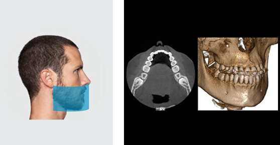
FOV 10X8cm
Perfect view of the two dental arches. The 10 cm diameter includes third molars, even in adult patients. Also available with 8 cm diameter for children and patients with a small build.
FOV 10X6cm
Complete dental arch in the adult. The 6 cm height combined with good positioning always ensures inclusion of all the necessary structures, without cutting out the occlusal zone or the base of the lower jaw.
FOV 8X6cm
Reduced view to examine a complete single dental arch in children or in patients with a small build.
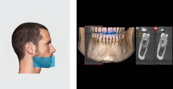
FOV 6X6cm
Ideal for sectional view along the dental arch. Partial scan of the individual semi-arches or frontal zone allows to considerably reduce the radiated dose.
Perfect practicality
- General dentistry
- Implantology
- Endodontics
- Gnathology
- General orthodontics
- Otorhinolaryngology
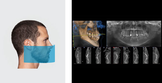
The ideal configuration for full dental applications, from endodontics to orthodontics and otorhinolaryngology (ENT). Using patented technology, GiANO HR Advanced can generate single volumes of up to 13 x 16 cm, providing a complete overview of dentition, maxillary sinuses and airways.
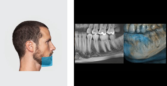
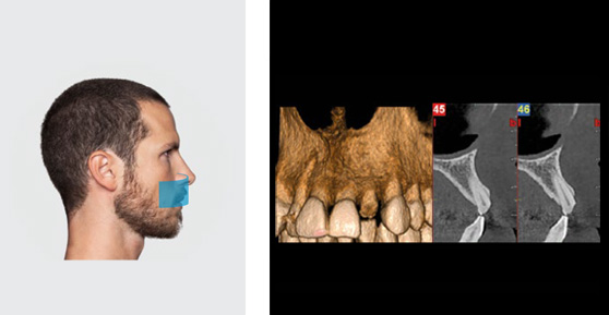
An examination performed with GiANO HR highlights characteristics, such as micro-fractures, bone height, root shape and tilt with the utmost precision.
Low X-ray doses, combined with the 3D aMAR (autoadaptive Metal Artifacts Reduction) function, illustrate anatomical structures clearly even in the presence of metallic objects. An essential requisite for post-surgery scans.
FOV 13X16cm
Single volume maxillofacial image obtained automatically: a complete view of the maxillary sinuses and of the entire dental arches. Also ideal for upper airway examinations.
FOV 10X10cm
For a complete examination of teeth, including the maxillary sinuses, with a single 360° scan and the option of an ultra rapid 6.4 s scan.
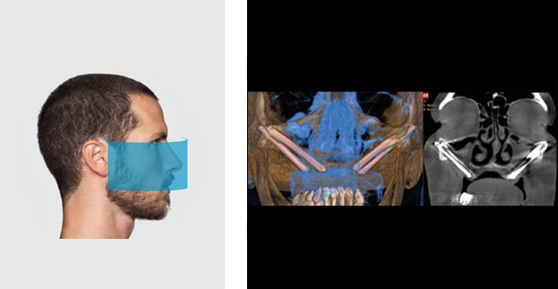
FOV 13x8cm
For wide range analysis of ascending rami of the mandible or of the zygomatic maxillary region for advanced implant design.
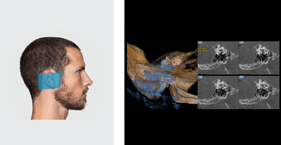
FOV 7x6cm
High definition image of both the internal ear and the petrous bones for accurate diagnosis or post-surgery control, such as positioning of the cochlear implant.
Absolutely perfect
- General dentistry
- Implantology
- Endodontics
- Gnathology
- General orthodontics
- Otorhinolaryngology
- Maxillofacial
- Head&Neck
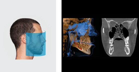
Sharp, detailed 3D images of the entire dental-maxillofacial area with a 16 x 18 cm volume at the highest resolution available on the market. Also useful for otorhinolaryngology investigations. Pathologies of the cervical spine can be studied using dedicated trajectories.
Examination of both temporomandibular joints in a single scan allows identification of any dysfunction based on the joint space image. Pre-set radiation levels and exclusive SafeBeam™ technology, available with all the configurations, lets users select the best exposure and obtain the optimal dose.
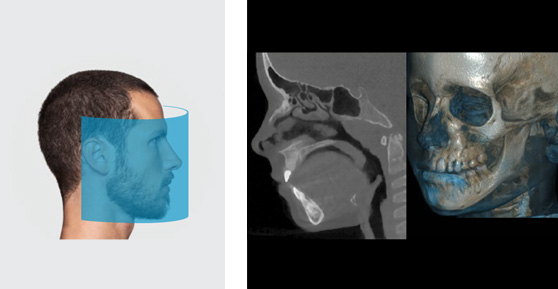
FOV 16X18cm
Scan view of upper airways from the nose to the trachea, both temporomandibular joints, maxillary and nasal sinuses.
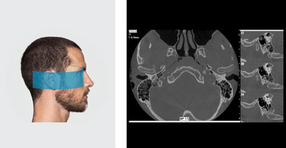
FOV 15X6cm
Detailed high resolution diagnosis of both temporomandibular joints or of the ear in a single scan.
FOV 16x10cm
Complete panoramic diagnosis of mouth, maxillary and mandibular structures up to the temporomandibular joints.
FOV 4x4cm
Limiting exposure to the region of interest and using 3D XF (eXtra Functions) allows to considerably reduce the radiated dose and to obtain images with exceptional resolution of 68 μm. For endodontic applications and optimal view of details, such as any secondary canals.