Ultra HD Imaging
DC TECHNOLOGY
Normal sensors are sensitive to visible light, and it is therefore necessary to convert X-rays through a scintillator so that they can be captured by the sensor. With DC technology, however, the sensor receives and processes X-rays directly, without any intermediate conversion. In this way, high resolution images are obtained with an excellent level of contrast, and with very low X-ray doses safeguarding patient health. Moreover, the sensor is stronger and less bulky, since unlike conventional technology intraoral sensors it does not contain traditionally fragile components.
MAXIMUM SHARPNESS
NewTom has always worked to offer the highest image quality standard for reliable diagnoses. DC technology applied to the sensor delivers X-ray imaging with a very high degree of detail, allowing dentists to carefully evaluate the resulting clinical picture.
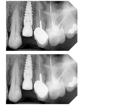
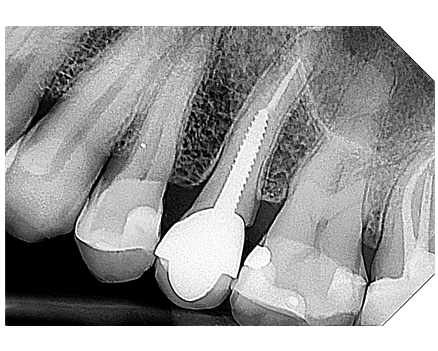
THE ADVANTAGES OF NEWTOM DCiS
A difference clearly emerges from the comparison between X-ray images obtained with traditional sensors and those obtained with direct conversion technology: NewTom DCiS dramatically improves contrast and sharpness enhancing the visibility of conditions that otherwise would not always be clearly diagnosable.
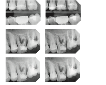
NEWTOM ADAPTIVE MULTIVISION
The NNT software provides the dentist with ApT (Adaptive picture Treatment) filters in Adaptive MultiVision mode and suitable for different clinical needs. You can select which filters to use from pre-set families or define customised ones based on individual diagnostic or visual preferences. In particular, it is possible to choose to preserve the visibility of soft tissues, keeping unaltered any areas at risk of image darkening. It is also possible to increase the contrast, if the desired level cannot be obtained for anatomical reasons, or due to the radiological parameters set. A default filter has the task of balancing noise, contrast and grey scale, while an additional filter enhances the details of the anatomical area examined. Finally and exclusively for the bitewing examinations, the caries revealing filter highlight the presence of interproximal caries.