.png)
.png)
Ergonomics
Newtom X-VS E VET is designed to optimise veterinary clinic workflows by ensuring excellent operational ergonomics.
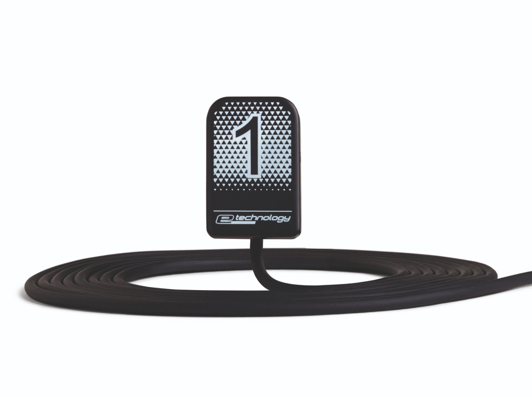
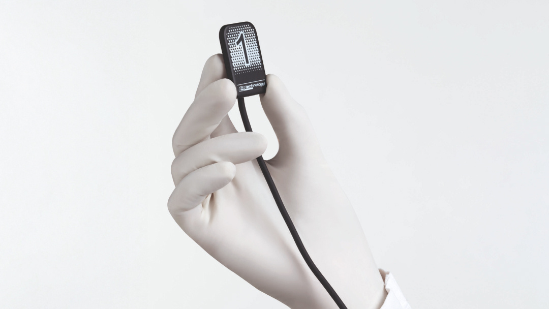
The ultra-thin sensor with rounded corners ensures quick and easy positioning, minimising animal discomfort. Thanks to the large active area and dedicated positioners, each exam is performed with outstanding precision and reliability.
The 3-metre flex cable ensures freedom of movement, allowing practitioners to work comfortably even where space is limited. Direct USB connectivity speeds up image acquisition and simplifies integration with existing systems.
The IP68-certified sensor is liquid- and dust-resistant, making it safe and hygienic. The NNT software enables the analysis and processing of images for diagnostic purposes or to help formulate a treatment plan.
X-VS E VET merges ergonomics and performance to deliver fast, precise diagnoses while ensuring comfort for both practitioner and animal.
Tools
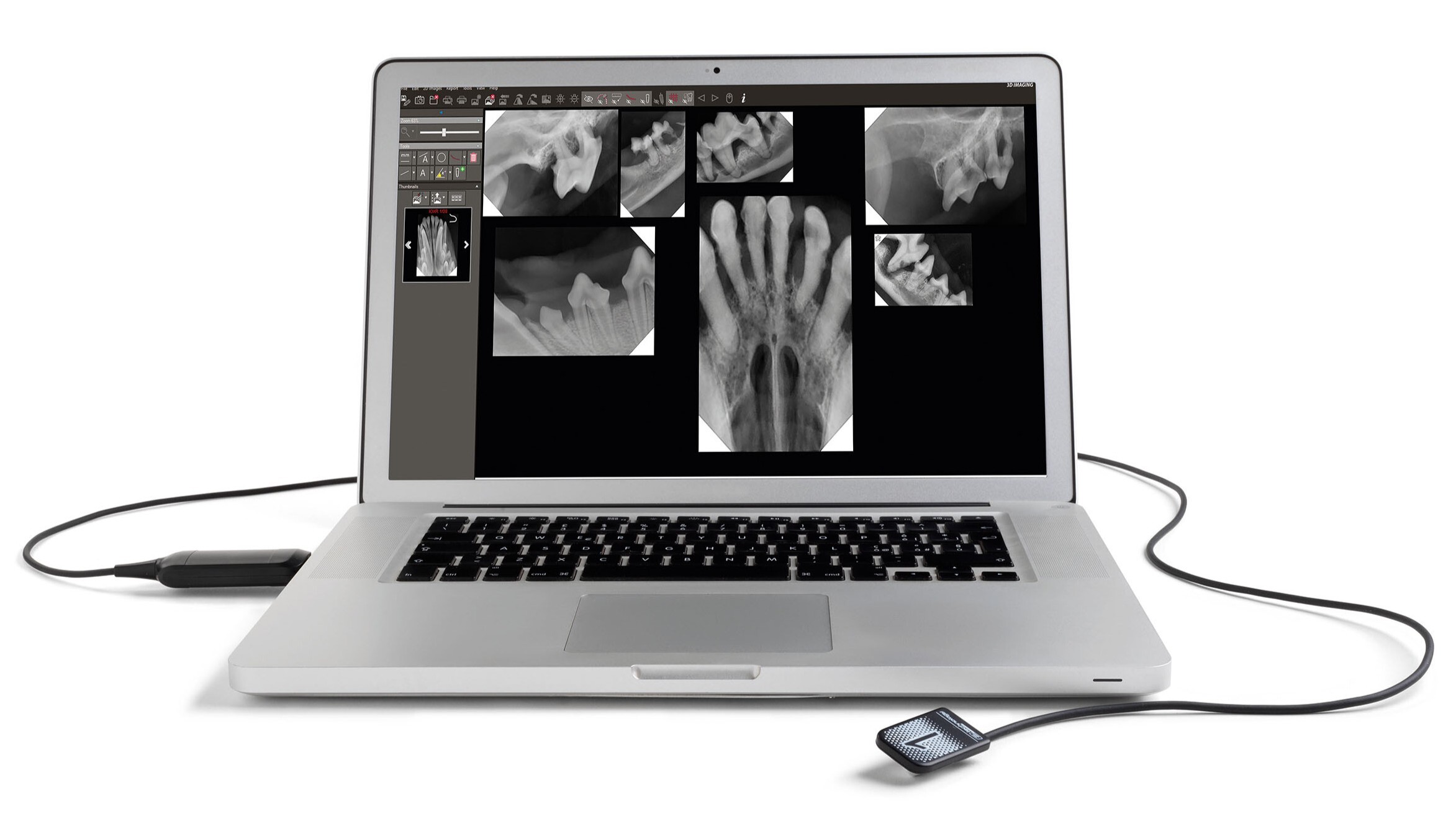
From the operator’s perspective, Newtom X-VS E VET is an effective tool that simplifies and optimises every stage of the X-ray scan. Dedicated positioners ensure correct placement of the sensor for different types of scan, minimising positioning times and maximising the quality of the images, which are loaded directly onto the PC and viewable immediately.
This makes X-VS E VET a powerful tool for quick and easy sharing, communication and storage, optimising the workflow.
Direct USB connectivity ensures fast, uninterrupted transfers, eliminating the need for intermediate devices. This reduces operational complexity, allowing users to focus exclusively on scan quality.
The NNT software has been developed to manage the acquisition of clinical images and organise them within the patient database. It enables the analysis and processing of images for diagnostic purposes or to help formulate a treatment plan, and also allows data and images to be shared with other complementary or alternative systems.
.png "Banner download Newtom (5)")